When you have something mysterious happening with your health, such as leg pain or unexplained swelling, a doctor can only see so much during an exam. Sometimes, they need assistance from technology to gain a deeper understanding of what’s happening inside your body. This is where diagnostic imaging comes into play.
You’re probably familiar with these tests by their names, like X-rays, MRIs, ultrasounds, and Doppler ultrasonography. Imaging such as ultrasounds helps healthcare providers make informed decisions, ultimately leading to better patient outcomes.
In this article, we’ll simplify the concept of diagnostic imaging, explaining how it works and the valuable insights it provides to patients and medical professionals. You’ll gain a comprehensive understanding of ultrasounds, why your doctor may recommend one, the various types available, and, most importantly, their profound significance in quicker and more accurate diagnosis.
Understanding Imaging and Ultrasounds
The easiest way to understand diagnostic imaging is to think of these tests as a portal into the inner workings of your body. Various forms of diagnostic imaging tests are available to capture these internal snapshots.
Common diagnostic imaging tests beyond X-rays include magnetic resonance imaging (MRI), computed tomography (CT) scans, and ultrasounds. The choice of test your doctor orders will depend on your specific symptoms. For instance, if there is a concern about a possible blood clot, your doctor will likely order a vascular ultrasound.
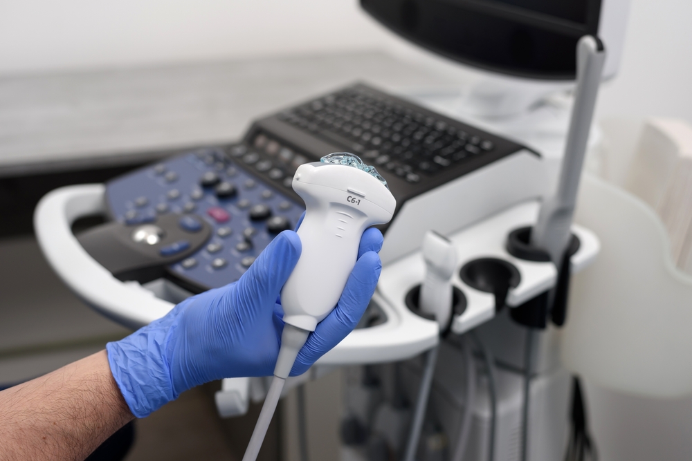
An ultrasound, often referred to as ultrasound imaging or sonography, is a diagnostic imaging technique that uses high-frequency sound waves to generate real-time images of structures within the body. Unlike X-ray imaging, ultrasounds do not involve exposure to harmful radiation. Instead, they rely on the principle of sound wave technology.
The specifics of how an ultrasound works depend on the ultrasound machine used, the medical context, and the purpose of the examination. But typically, a device called a transducer emits these high-frequency sound waves, which then bounce off internal structures and return as echoes. By analyzing the time it takes for these echoes to return and their intensity, the ultrasound machine creates detailed visual representations of the targeted area.
These internal images, generated through sound wave technology, provide your doctor with the insights necessary to thoroughly evaluate your condition and make precise diagnoses, all while ensuring patient safety by avoiding excess radiation exposure.
The Importance of Imaging and Ultrasounds
Imagine testing such as ultrasounds plays a pivotal role in enabling healthcare providers to visualize the body’s inner workings, which is crucial for accurate medical diagnosis.
Among them, ultrasounds and Doppler ultrasounds are the most widely used and preferred imaging tests by both patients and medical professionals, mainly due to their versatility and non-invasive nature.
Ultrasounds are considered necessary in the context of healthcare for numerous reasons:
Diagnostic Accuracy
Ultrasounds significantly enhance diagnostic accuracy. By creating detailed images of internal structures, they enable healthcare professionals to identify problems that might be challenging to detect through physical examinations alone. This capability is particularly vital for early and precise diagnosis, ensuring that patients receive appropriate treatment promptly.
Real-time Imaging
Ultrasounds provide real-time imaging, allowing healthcare providers to observe the body’s internal dynamics as they happen. One notable example of its significance is in vascular health assessments.
During a vascular ultrasound, clinicians can monitor blood flow in real-time through arteries and veins. This immediate feedback is invaluable when timely decisions about circulatory health and potential blockages are critical, enabling prompt intervention to safeguard a patient’s well-being.
Non-Invasiveness and Patient Safety
Ultrasounds are non-invasive and pose no risk of ionizing radiation exposure. This makes them an important tool in healthcare, particularly for vulnerable patient populations, such as pregnant women, infants, and those with contraindications to other imaging methods.
Cost-Efficiency
Ultrasounds are a cost-effective imaging solution compared to alternatives such as MRIs or CT scans. Insurance typically covers ultrasounds, but their affordability extends beyond insurance coverage. This accessibility ensures that even for patients without insurance, these frequently essential tests remain a viable and practical option for their healthcare needs.
Why You Might Need an Ultrasound
Ultrasounds are instrumental in diagnosing various medical conditions and symptoms. There are numerous reasons why an ultrasound may be ordered by your healthcare provider. While pregnancy is a well-known application, it only scratches the surface of its versatility.
Here are some reasons why your doctor might order an ultrasound:
Vascular Health
Vascular ultrasounds are pivotal in assessing blood flow and identifying issues such as blood clots, artery blockages, or aneurysms. They are vital for diagnosing conditions like deep vein thrombosis (DVT), peripheral artery disease (PAD), and carotid artery disease.
General Health
“General” ultrasounds can assess various organs, including the thyroid, pancreas, and spleen. They are valuable for uncovering abnormalities, cysts, or tumors within these structures.
Plastic and Surgical Oncology
Ultrasound imaging has also found its way into plastic and surgical oncology. In plastic surgery, preoperative ultrasounds enable precise planning for reconstructive procedures, optimizing surgical design and dissection techniques. In surgical oncology, ultrasound-guided procedures aid in the localization of tumors, ensuring targeted and effective treatment.
Abdominal Disorders
Ultrasounds of the abdomen assist in diagnosing gastrointestinal issues, gallstones, liver disorders, and kidney conditions. They offer a non-invasive means of investigating abdominal pain, digestive problems, or unexplained symptoms.
Cardiac Evaluation
Echocardiograms, a specialized form of ultrasound, are employed to evaluate heart function, detect heart valve abnormalities, and diagnose congenital heart conditions. They play a critical role in cardiology.
Types of Ultrasounds
When most hear the term “ultrasound,” the first image that often comes to mind is a pregnant mother’s ultrasound or a breast ultrasound to help diagnose breast cancer. However, medical imaging goes beyond “breasts and babies,” offering many possibilities.
Here are some of the most common types of ultrasounds and ultrasound techniques used to diagnose a host of medical issues:
1: Vascular System Ultrasounds and Imaging
Vascular ultrasounds are pivotal in assessing blood flow and diagnosing vascular conditions. They offer a window into the circulatory system, ensuring early detection and intervention when necessary. There are numerous imaging tests or diagnostic procedures that involve the use of ultrasound or Doppler techniques to visualize and assess blood vessels and circulation.
Here is a comprehensive list of vascular imaging and ultrasound exams that focus on assessing blood vessels and circulation:
Arterial Doppler with Treadmill (Exercise Doppler)
This specialized ultrasound assesses blood flow in arteries, especially in the lower extremities, measuring changes in blood flow and velocity in response to physical activity. This can reveal circulatory abnormalities that may not be apparent at rest. It assists in diagnosing vascular diseases and peripheral artery issues, ensuring early intervention when needed.
Venous Reflux
Venous reflux exams primarily focus on the venous system, specifically the veins. These tests are designed to detect and assess abnormalities in venous blood flow, such as venous insufficiency or varicose veins.
Venous Duplex Post EVLT
This exam evaluates the status of veins in the venous system, particularly after undergoing Endovenous Laser Treatment (EVLT) for varicose veins. It ensures that the treated veins remain functional and unobstructed.
IVC and Iliac Vein Duplex
IVC and iliac vein duplex exams concentrate on the inferior vena cava (IVC) and iliac veins, essential components of the venous system. These tests are performed to identify blockages, narrowing, or other abnormalities that may affect the blood flow from the lower body to the heart.
Upper and Lower Extremity Arterial and Venous Duplex
These exams thoroughly assess the arterial and venous systems in the upper and lower limbs. They are designed to detect conditions such as arterial stenosis or venous insufficiency in the arms and legs.
Aorta Iliac Duplex
The focus of this exam is the aorta and its branches, particularly the iliac arteries. It aims to identify and assess aortic aneurysms, stenosis, or other abnormalities that may affect the main arterial pathways.
Aorta and Aorta Endograft Duplex
These exams monitor the condition of the aorta, especially when an endovascular graft has been placed. They ensure the proper functioning of the graft and detect potential complications that may arise.
Dialysis Access Duplex
Dialysis access duplex exams evaluate the patency and functionality of blood vessels used for dialysis access, such as arteriovenous fistulas or grafts, which are essential for efficient dialysis treatment.
Mesenteric and Renal Artery Duplex
These exams specifically focus on the mesenteric and renal arteries, vital for blood supply to the intestines and kidneys, respectively. They aim to detect stenosis or other vascular problems that may impact blood flow to these organs.
Carotid Duplex
Carotid duplex exams target the carotid arteries in the neck. They are performed to identify plaque buildup or stenosis in the carotid arteries, conditions that increase the risk of stroke.
Bypass Graft Duplex
These exams assess the patency and function of vascular bypass grafts that reroute blood flow around blocked arteries, examining blood vessels with bypass grafts.
Vein Mapping
Vein mapping is often conducted on the veins in the legs. It is typically done in preparation for procedures that require healthy veins, such as coronary artery bypass surgery.
ABI (Ankle/Brachial Index)
The ABI exam evaluates the arterial system in the arms and legs. It involves measuring blood pressure to diagnose peripheral arterial disease (PAD) by comparing blood pressure measurements in the ankles and arms.
Upper and Lower Extremity Doppler
These exams investigate the arterial and venous systems in the upper and lower limbs. They use Doppler ultrasound to measure blood flow velocity and detect conditions like arterial stenosis or venous insufficiency.
2: General Ultrasounds and Imaging
General ultrasounds are versatile tools that enable detailed examinations of various organs. They’re instrumental in diagnosing a wide range of medical conditions, from abdominal issues to thyroid disorders.
Here are a few of the most common general ultrasounds:
Gallbladder Ultrasound
By examining the gallbladder, we can identify issues like gallstones or other abnormalities that might be causing discomfort or digestive problems.
Renal Ultrasound
Focusing on the kidneys, this ultrasound helps us evaluate their size, shape, and overall health. It’s an essential tool in diagnosing kidney conditions.
Thyroid Ultrasound
If you’re experiencing thyroid issues, this ultrasound allows us to examine the thyroid gland for nodules or other thyroid disorders.
Abdominal Ultrasound
This ultrasound provides detailed imaging for a comprehensive view of your abdominal organs. It’s an invaluable resource for diagnosing gastrointestinal issues and other abdominal conditions.
3: Plastics and Surgical Oncology Ultrasounds
In the fields of plastics and surgical oncology, precision is paramount. Ultrasounds are often used to guide procedures and assist in precisely locating tumors or lesions, ensuring targeted tissue sampling during biopsies (ultrasound-guided biopsy.) This level of accuracy is essential for planning successful surgeries and treatments.
4: Breast Ultrasound
Breast ultrasounds are designed explicitly for evaluating breast health. They assist in detecting subtle abnormalities within breast tissue, making them essential for early detection.
While they are not a substitute for mammograms, they complement these screenings effectively. When a patient presents abnormal mammogram results, breast ultrasounds can provide a comprehensive evaluation.
Moreover, as previously mentioned, they play an essential role in oncology, guiding biopsies and ensuring precise interventions when needed.
What to Expect During Imaging and an Ultrasound
The exact method and steps during diagnostic imaging can vary based on the type of imaging or ultrasound you are having performed. However, generally, an ultrasound is a straightforward procedure.
You’ll receive specific guidelines from your healthcare provider, which may include wearing comfortable clothing and avoiding lotions or creams on the area to be examined. If there are dietary or hydration requirements, these will be communicated to you in advance.
During the procedure, you’ll lie on an examination table, and a skilled technician will use a small, hand-held device called a transducer. This device emits high-frequency sound waves into your body, creating real-time images on a monitor. It’s a painless process; the only thing you might feel is slight pressure.
Once the ultrasound is complete, you can resume your normal activities immediately. There’s typically no downtime or recovery needed. Your healthcare provider will review the images and discuss the findings during a follow-up appointment.
Why Medical Ultrasound Awareness is Important
Ultrasound imaging is a crucial diagnostic tool that provides precise insights capable of saving lives. Promoting awareness about these tests is vital, allowing individuals to grasp their importance in healthcare. Moreover, it’s an opportunity to applaud the skilled professionals who use ultrasound to enhance patient care.
In October, we observe Ultrasound Awareness Month, a dedicated time to underscore the significance of this incredible imaging tool in our journey through the intricate world of modern healthcare.
You can participate in Ultrasound Awareness Month by supporting local awareness events, sharing information with friends and family, and most importantly, scheduling an ultrasound if your doctor has suggested that you might need one.
Where to Book a Vascular Ultrasound or General Ultrasound in Middle Tennessee
With The Surgical Clinic, you have a trusted partner for vascular and general ultrasounds in Middle Tennessee. Our labs are equipped with Registered Vascular Technologists (RVTs) holding RVT certification, ensuring your tests are conducted by skilled professionals.
TSC Vascular Labs
Our labs are accredited by the Intersocietal Accreditation Commission, and our RVTs are registered through the American Registry of Diagnostic Medical Sonographers (ARDMS). These labs play a pivotal role in providing accurate diagnoses and effective treatment options for vascular conditions.
TSC General Ultrasounds
Our general ultrasounds employ advanced imaging technology that utilizes high-frequency sound waves to create real-time images of the inside of your body. We offer three ultrasounds types: conventional, three-dimensional (3-D), and four-dimensional (4-D) options.
For your convenience, we offer our imaging services in multiple Middle Tennessee locations. We even provide ‘same-day’ appointments, and your test results are promptly shared with your surgeon to determine the best course of treatment.
We also offer low-cost vascular health screenings at our vascular laboratory that help with early detection of the most common types of vascular disease. Vascular screenings help catch any irregularities and prevent issues before you even need a vascular ultrasound. We can help you find out if you are at risk and who should get a screening. Contact us today for more information on booking at an imaging center near you.