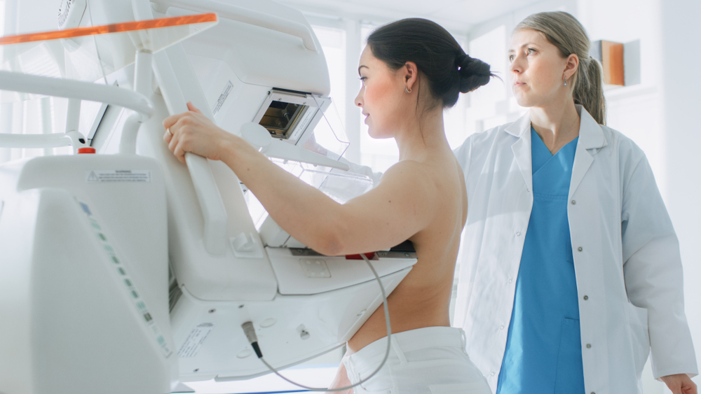
New technologies have given patients many choices about what types of mammograms to have and the difference between the types of studies can be confusing. As a surgeon at The Surgical Clinic who specializes in Breast Surgery, I am often asked what the difference between these exams is and why patients will be asked to have different types of studies as we decide together the best approach to treatment.
A “screening” mammogram is just that, a method to screen the breast for abnormalities such as calcifications, masses, or areas of distortion. If identified, a screening mammogram will transition into a diagnostic” mammogram. A diagnostic mammogram means that an abnormality has been identified requiring further imaging in an attempt to diagnose the area(s) in question. Although the timing and need for screening mammograms has recently come under attack, again. This info-blog will concentrate on the types of screening mammograms available and some of the misconceptions. I supply this information to my patients for their education. This is not intended to be a comprehensive review, but an overview of the methods for screening mammography.
Xeromammography was developed in the 1960’s and first used to image soft tissue later being adapted to detect breast cancer. This type of mammogram can be identified by the baby blue color and appears as a “negative” mammogram. Used from 1965-1975, it was welcomed by women because of the slight compression used during the process. This slight compression was the Achilles heel of the procedure and did not allow for visualization of the posterior breast or chest wall. So, due to the inability to visualize the entire breast, xeromammography fell by the wayside. Today, dedicated breast centers are not performing this type of screening mammograms.
Thermomammography is the process of using an infrared camera to produce an image of the heat emitted by the breast. The heat signature is directly proportional to blood flow. The FDA first approved the technology in 1982 as an adjunct (additional test) for detecting breast cancer. Studies have shown that thermography is not an acceptable method of screening the breast. This is the current position of the FDA, Susan G. Komen Foundation and American Cancer Society. Free standing thermography centers have opened across the country making false claims regarding the use of screening thermography. This has sparked “Warning Letters” from the FDA to these centers. Canada went so far as to call the procedure a sham! Thermography is not a substitute for x-rays and is not recommended for screening.
Analog Mammography was the first method used for screening. This is analogous to the old analog cameras that exposed an image onto a film requiring development before the image could be seen resulting in reading delays. The size of the film dictated the size of the breast which could be imaged. Two images were taken. One image was the CC (craniocaudal) view looking down on the breast from the head to feet. The second image was the MLO (medial to lateral oblique) looking across the breast from the breast bone to the arm at a slight angle to incorporate the breast tissue under the arm. These two images are called 2-D or 2 Dimensional. Archiving and transferring this type of image is much more difficult. Converting analog to digital is at best, very difficult. Reviewing this type of image was traditionally done with a magnifying glass! Just like analog cameras gave way to digital cameras, so too did analog mammography.
Digital Mammography has been dubbed the latest medical “darling”. This method is like the digital camera on your phone giving the radiologist the tools needed to manipulate the images. The two views (CC and MLO) are still obtained, but the delay in reading is significantly less as the images are available immediately. This is still a 2-D method of looking at the breast. Archiving and transferring images are as simple as a click of the button. After the mammogram, I can review the images in my office with the patient. Some say that digital mammograms detects cancer earlier than analog, but after 22 years of breast surgery, my opinion is that it’s the experience of the reader. As good as digital mammography is, we made it better!
Digital Mammography with 3-D Tomosynthesis uses the conventional 2-D images, but adds 11-15 images taken across a 15o arc of the breast. The example is a bird in a tree. If you look at the tree from 2 views which are perpendicular, the bird may not be seen. If you look at the tree from 2 perpendicular views and walk in a 15° arc around the tree, you will see the bird much better How does 3-D compare to 2-D breast imaging? The recall rate for a 3-year period for 2-D imaging was 10.4% while the rate for 3-D imaging was 9.0%. The rate of cancer detection in women who were recalled was 4.4% in 2-D and 6.5% in 3-D. Finally, in women who consecutively underwent 3-D mammography the recall rate declined every year from 13% in year one to 5.9% in year 3. So, the rate of recall is less in women who have 3-D imaging, the rate of cancer detection is higher and with consecutive 3-D mammograms the recall rate decreases. This decrease in recall rate addresses the current controversy regarding unnecessary mammograms. One caveat to your choice in screening mammograms is that not all insurance companies cover tomosynthesis leaving the patient with an out of pocket expense. Tomosynthesis can save patients from having a screening mammogram converted into a diagnostic exam.
As a breast surgeon, tomosynthesis is a welcomed addition to imaging the breast. It can decrease recall rates and unnecessary biopsies. I hope this has been useful to your choice in screening mammography.

